The Heart is the key to the world and to Life.
We live in our present helpless condition
In order to love one another
And be obliged to help one another.
Through imperfection we become open
To the influence of others
And this influence from outside is the aim
That in our frailties
Others can and may help us.
—Novalis
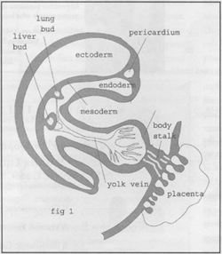 In the embryology of the human heart the first forms that
appear reveal a deep mystery to physiologists. The middle layer, or mesoderm,
is the most active of the three layers in the early embryo. It proliferates
inside and even outside of the embryo early in the first week. At that time, an
inner space mysteriously opens in the mesoderm. (see fig.1) This magical space,
or pericardium, appears, with no previous indicators, just outside of the head
on the periphery of the embryonic disc. The pericardium then enters through the
primal mouth opening of the inner mesoderm and descends through the body of the
embryo towards the chest. Simultaneously, near the tail end, other mesodermal
cells modify into blood-filled veins known as yolk veins. The pericardial cavity
and the yolk veins develop on opposite ends, outside of the body of the embryo.
Eventually the yolk veins from below will meet the descending pericardial space
and penetrate it in the very center of the body. The union of these two polar
developments is the formal motif that underlies the miracle of the human heart.
In the embryology of the human heart the first forms that
appear reveal a deep mystery to physiologists. The middle layer, or mesoderm,
is the most active of the three layers in the early embryo. It proliferates
inside and even outside of the embryo early in the first week. At that time, an
inner space mysteriously opens in the mesoderm. (see fig.1) This magical space,
or pericardium, appears, with no previous indicators, just outside of the head
on the periphery of the embryonic disc. The pericardium then enters through the
primal mouth opening of the inner mesoderm and descends through the body of the
embryo towards the chest. Simultaneously, near the tail end, other mesodermal
cells modify into blood-filled veins known as yolk veins. The pericardial cavity
and the yolk veins develop on opposite ends, outside of the body of the embryo.
Eventually the yolk veins from below will meet the descending pericardial space
and penetrate it in the very center of the body. The union of these two polar
developments is the formal motif that underlies the miracle of the human heart.
The mesoderm completely permeates the inside and the outside
of the embryo in its early development. Buds of organs rise out of the mesoderm like buds rising out of
the cambium of a tree. Some buds are formed outside of the embryo and some are
within the embryo. (see fig.2)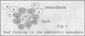 In the first week many of these buds form in the
mesoderm of the yolk sac in the gut region of the embryonic disc. In these yolk
sac buds, the cells that are on the periphery of the bud flatten and form
plates. (see fig.3) At the same time, cells in the center of the yolk sac buds
form donut-shaped blood cells.
In the first week many of these buds form in the
mesoderm of the yolk sac in the gut region of the embryonic disc. In these yolk
sac buds, the cells that are on the periphery of the bud flatten and form
plates. (see fig.3) At the same time, cells in the center of the yolk sac buds
form donut-shaped blood cells.
As the buds proliferate on the periphery of the yolk sac, the flattened plates
of cells join each other on their sides and then canalize into each other to
form islands of capillaries.
Inside the canals of the capillaries, the donut-shaped blood cells from the
center of the buds form the blood itself. Together the vessels and blood cells
form vascular, sponge-like tissues called blood islands. These blood islands
are the first seed-like formation of the heart in the metabolic region.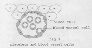
It is hoped that with these imaginations a spark of interest
can be kindled to study the morphology of our human body as a central motif in
the task of self-knowledge. Physiology and morphology are the keys to the
secret door of self-knowledge. When we can picture inwardly the sequences of
creative movements that the Hierarchies have presented to us in the sublime
morphology of the embryo, the door to self-knowledge is unlocked. Meditatively
working with the images as a sequence of inner pictures slowly opens the inner
door to an experience of the imaginative capacity in the soul. In this way the
embryo can be experienced as the keeper of the keys to self-knowledge.
Dennis Klocek, Director of the Consciousness Studies
Program at Rudolf Steiner College, Fair Oaks, CA; author of several books and
international lecturer. One of the keynote presenters with Bert Chase,
architect, in the conference at RSC June 16-20, Embryology, Architecture and
the Origins of Form.